
Cell biology template
Cell Membrane Diagram
Build a labeled cell membrane diagram for teaching, research figures, pathway explanations, or a graphical abstract.
Create a membrane diagram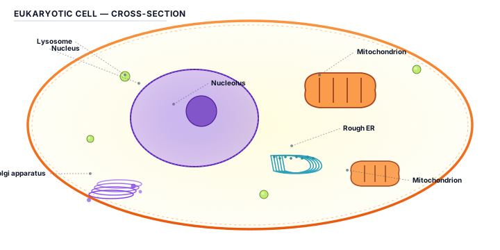
Cell biology template
Build a labeled cell membrane diagram for teaching, research figures, pathway explanations, or a graphical abstract.
Create a membrane diagramA useful cell membrane diagram makes the boundary of the cell easy to recognize and then explains what crosses it or interacts with it. Depending on the research question, the visual may include the phospholipid bilayer, membrane proteins, channels, transporters, receptors, extracellular ligands, cytoplasmic factors, or a signal moving toward the nucleus. The diagram does not need to show every molecule. It should show the parts that support the claim and use labels that match the terminology in the paper or lesson.
Use a cross-section when the goal is to explain layers and transport. Use a pathway layout when the membrane is the starting point of a signaling cascade. A central membrane with outward callouts works well for a labeled educational figure. SciDrawer can help create a starting composition from a text prompt or sketch; check orientation, compartment names, and the direction of every arrow before using it in a formal figure.
Start by deciding whether the diagram explains structure, transport, receptor activation, or a comparison between membrane states. Name the compartments first, then list only the proteins and molecules needed for that explanation. Keep the bilayer visually stable so the reader can distinguish the extracellular and intracellular sides at a glance.
Use short labels for membrane components and add a compact legend for repeated symbols. If the figure includes a receptor or transporter, state what it binds or moves. The plant cell labeled example shows how a stable central structure and outward leader lines can keep many labels readable.
A receptor becomes meaningful when the diagram shows ligand binding, conformational change, or a downstream signal. A channel becomes meaningful when an ion or molecule has a clear direction. Use arrows and annotations to explain the relationship, then remove decorative elements that compete with the process.
Compare the final labels with the manuscript or source material, test the diagram at the target display size, and export the format required by the journal or course. If the diagram becomes part of a paper-wide visual story, compare it with the scientific diagram maker and graphical abstract maker workflows.
Avoid mixing the two sides of the membrane without a clear compartment label. Do not draw arrows through the bilayer if the process is meant to stop at the membrane, and do not use color alone to distinguish extracellular from intracellular events. Labels should not cross the membrane or each other, and abbreviations should be explained when the figure is intended for a broad audience.
Yes. Describe the bilayer, compartments, proteins, molecules, and labels you need, then review the generated structure for scientific accuracy.
Yes. Specify the direction, starting event, and outcome so the diagram explains a relationship rather than showing isolated objects.
Yes. A sketch is useful for communicating the layout and the relative position of compartments or membrane components.
No. Check every label, arrow, compartment, and biological claim against your manuscript or source material before publication.
Yes. Replace the labels with the preferred Chinese terminology and check font support and readability for the final audience.
Describe the membrane structure or process you need, attach a sketch if useful, and create a scientific starting point with SciDrawer.
Open the AI figure generatorExplore more focused research figure pages, browse editable templates, or open the generator for a custom draft.